
Some critical notes on “Comment on ‘Triassic coleoid beaks and other structures from the Calcareous Alps revisited’ by Doguzhaeva et al. (2022)” by Lukeneder and Lukeneder (2022)”
LARISA A. DOGUZHAEVA
The compact micro-laminated ultrastructure of the undeniable beak of the Carnian (Late Triassic) Lunzoteuthis schindelbergensis Doguzhaeva, Summesberger, and Mutvei, 2006, from the Calcareous Alps, that was buried together with the Phragmoteuthis bisinuata (Bronn, 1859), showed fine fossilization of chitin (Doguzhaeva et al. 2006; Doguzhaeva and Summesberger 2012). Hence, the hypothesized “beak of P. bisinuata” (Suess 1865; Mojsisovics 1882; Jeletzky 1966; Rieber 1970) should have, similarly to the beak of L. schindelbergensis, a chitinous composition. However, this “beak” revealed “a vertebrate cartilage inducing a bone growth” (Fig. 1) meaning a fragment a juvenile vertebrate, that might be a P. bisinuata’s catch (Doguzhaeva et al. 2022: 9).
The “beak of P. bisinuata” hypothesis is, therefore, rejected on the basis of the cartilaginous composition of the “beak of P. bisinuata”, and the SEM images represent the main factual basis of the paper by Doguzhaeva et al. (2022). Hence, a criticism of this paper requests consideration of the SEM images evidencing “a vertebrate cartilage inducing a bone growth”. However, Lukeneder and Lukeneder (2022) do not discuss the “factual basis” (= SEM data) of the paper commented on by them (Doguzhaeva et al. 2022), and a key information on complex bone nanostructures in the matrix of the cartilage of the “beak of P. bisinuata” (Fig. 1) incompatible with the collagen fibers of the cranial capsule, is not considered by them. Hence, the “Discussion by Lukeneder and Lukeneder (2022)” represents a personal opinion that does not stem from the analysis of the “factual basis” of the commented paper (Doguzhaeva et al. 2022).
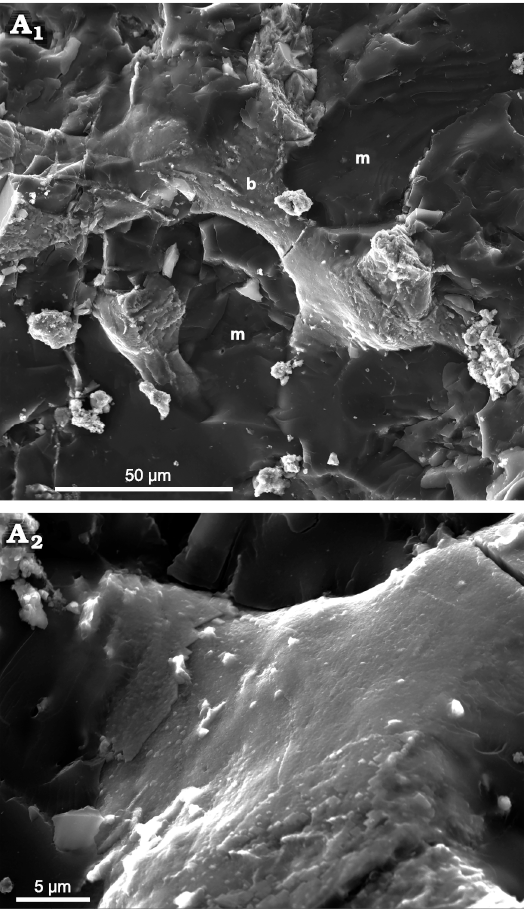
Fig. 1. A fracture of the hypothesized “beak” of the coleoid cephalopod Phragmoteuthis bisinuata (Bronn, 1859) (GBW 2006/011/0009); lower Carnian, Upper Triassic, Cave del Predil, NE Italy. A1, “glassy” (dark grey) structureless carbonized matrix (m) containing a whitish complex calcified nanobone (b); A2, enlargements showing a dense smooth (slightly dimpled) bone surface. GBA, Geological Survey of Austria, Vienna.
One of “the arguments against the ‘prey’ hypothesis” stated in Lukeneder and Lukeneder (2022: 965) is: “… the position of the preyed fish is variable, but prey remains in the stomach, beak, and arm crown are to our best knowledge unknown neither from Polzberg nor from Cave del Predil.” This is, however, not an argument against recent interpretation of the “beak of P. bisinuata”, but a constatation of the recent status of knowledge on fish taphonomy in the two localities. The fact, which is needed to be added here, is that Triassic deposits worldwide yielded so far, a sole coleoid showing a stomach content and it is the Early Triassic Idahoteuthis parisiana Doguzhaeva and Brayard, 2018, from Idaho, USA (Doguzhaeva et al. 2018).
Lukeneder and Lukeneder (2022: 964) “…are convinced that this idea of prey in the mouth of almost every studied specimen is erroneous and that the arguments of Doguzhaeva et al. (2022) can easily be falsified.” This sounds inappropriate, if not to say insulting. Do the authors believe that their older colleagues from the Natural History Museum of Vienna are able to falsify the data obtained with SEM (Doguzhaeva et al. 2006, 2007, 2010, 2022)?
Lukeneder and Lukeneder (2022: 963) inform that the “beaks” are “regularly occurring…”. However, this is an incorrect information as the potential “beaks” were preserved only in seven specimens from about 150 specimens studied by us (ca. 5%).
Lukeneder and Lukeneder (2022: 964) contains the statement: “These morphological consistencies convinced Lukeneder and Lukeneder (2022) to recognize the black structures as the cephalic cartilages rather than fish bones as Doguzhaeva et al. (2022) postulated (or beaks as Suess [1865] originally suggested).” The erroneous authors’ logic is here demonstrated with the aid of the specimen from Cave del Predil, NE Italy (Fig. 2), that, like a cranial capsule, has a pair of intact rounded “eye sockets” (Fig. 2). This specimen was found by the author in the shale with P. bisinuata in 2010. If to follow the above statement, it would be interpreted as a cranial capsule of P. bisinuata. However, the specimen bears numerous conodonts, evidencing that this is a conodontophorid.
Lukeneder and Lukeneder (2022: 964) unnecessarily freely replace some concepts with others. For example, “the cartilage inducing the growth of bones” of Doguzhaeva et al. (2022) is erroneously replaced with the “fish bones”. This is an unfair mistake that might be originated from the fact that the discovery of the cartilaginous tissue of black structures, associated with P. bisinuata, was made before the two authors of Lukeneder and Lukeneder (2022) began their study on this subject. The first SEM images of the cartilaginous ultrastructure of back structures associated with P. bisinuata were taken by my colleagues and me, labeled as “Cart” (cartilage) and “Ceph capsule” (cephalic capsule), and shown to the junior author of Lukeneder and Lukeneder (2022). Given the significant value of these rare data, the open access to these SEM images has been left by us for the scientific staff of the Department of Geology and Palaeontology of the Museum of Natural History of Vienna.
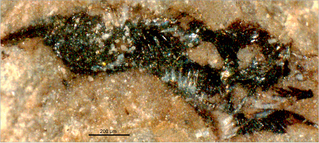
Fig. 2. A black, apparently cartilage structure of a conodontophorid, saving two intact “eye sockets” and numerous conodonts (NHMW 2012/0117/0023); lower Carnian, Upper Triassic, Cave del Predil, NE Italy. NHMW, Museum of Natural History of Vienna, Austria.
The final statement of the “Comment…” generates the following critical remarks: (i) “…the black carbonized structures are exceptionally located in the head region (never within the arm crown) ...”. The erroneous nature of this statement is evidenced by the fact that black structures can be preserved not only within the head area, but also outside it, and they can be surrounded by the arm hooks that are the elements of the arm crown (Doguzhaeva et al. 2022: fig. 4); (ii) “… the black carbonized structures … exhibit a consistent morphology”. This statement is disproved by the black structures shown in Doguzhaeva et al. (2022) and Fig. 2 in this paper. They can be the cartilaginous tissues of the conodontophorids; (iii) the authors “…could not find any evidence that support the prey idea of Doguzhaeva et al. (2022)”. As it was shown above, Lukeneder and Lukeneder (2022) lacks the discussion of the “factual basis” (= SEM data) of the paper by Doguzhaeva et al. (2022) and does not present any fact refuting the contested point of view. However, the difference between the bone nanostructures in the matrix of cartilaginous “beak of P. bisinuata” (Fig. 1) and Doguzhaeva et al. (2022: figs. 6A1, 8A1–A4, 9A1–A4, 10A1–A4, 11A1–A3) and collagen fibers of the cartilaginous cranial capsule of Loligo (Doguzhaeva et al. 2022: fig. 12C, D) is remarkable. This shows that the authors prefer not to look at the facts; (iv) the authors “… ascertain that they recorded the first Triassic cephalic cartilages”. This phase sounds good if one does not know that the labels CART1-CART28 and Ceph capsule 2010 used for SEM images taken by Doguzhaeva with the co-authors, were known to the second author a long time ago. One of these images was demonstrated in a poster session in the 3rd International Palaeontological Congress in London (Doguzhaeva et al. 2010). Therefore, they should have accompanied this phrase with gratitude for the fact that they started their research, thanks to our openness and that the discovery of the cartilaginous tissues of the black structures from the Carnian deposits of the Alps belongs to the authors of the paper commented on by them (Doguzhaeva et al. 2022). It should be noted that the coleoid affiliation of the oldest so far recorded Carboniferous cephalic cartilage is based on its association with a coleoid type radula and arm hooks (Doguzhaeva et al. 2010). Therefore, the morphological similarity alone leaves room for doubt (see Fig. 2); (v) “… the prey idea of Doguzhaeva et al. (2022)”. This expression incorrectly conveys the main idea of the paper by Doguzhaeva et al. (2022); the latter is focused on the problem of hypothesized “beak of P. bisinuata”.
In Lukeneder and Lukeneder (2022: 964), the inadequate testing of the described material, illustrated in Doguzhaeva et al. (2022: fig. 2B), resulted in an incorrect interpretation of the gladius associated with the arm crown and wrong conclusion that “… Doguzhaeva et al. proposed two different interpretations (Doguzhaeva et al. 2022: fish in fig. 2A and squid in fig. 2B) for one and the same structure, a fact that fundamentally mistrusts their prey idea”. The two different structures, showing different morphology, are considered by Lukeneder and Lukeneder (2022) as one and the same structure. The gladius associate with the imprints of the arm crown evidences a cannibalism of P. bisinuata that is not noted in Lukeneder and Lukeneder (2022), although this is only the second recorded fossil specimen showing this phenomenon (see Doguzhaeva et al. 2018).
Additionally, the interpretation of the cartilaginous “beak of P. bisinuata” as a cartilaginous piece of a vertebrate prey cannot be rejected without representing similar sort of the morphological, ultrastructural and evolutionary morphology data which would prove our observations to be wrong. Such information is missing in the commented paper.
Acknowledgements.—I would like to thank Editor-in-Chief Andrzej Kaim, who kindly invited me to respond to the Discussion by Lukeneder and Lukeneder (2022).
References
Doguzhaeva, L.A. and Summesberger, H. 2012. Pro-ostraca of Triassic belemnoids (Cephalopoda) from Northern Calcareous Alps, with observations on their mode of preservation in an environment of northern Tethys which allowed for carbonization of non-biomineralized structures. Neues Jahrbuch für Geologie und Paläontologie 266: 31–38. Crossref
Doguzhaeva, L.A., Brayard, A., Goudemand, N., Krumenacker, L.J., Jenks, J.F., Bylund, K.G., Fara, E., Olivier, N., Vennin, E., and Escarguel, G. 2018. An Early Triassic gladius associated with soft tissue remains from Idaho, USA—a squid-like coleoid cephalopod at the onset of Mesozoic Era. Acta Palaeontologica Polonica 63: 341–355. Crossref
Doguzhaeva, L., Mapes, R.H., Bengtson, S., Summesberger, H., and Meléndez Hevia, G. 2010. Ink, soft tissues and non-mineralized structures in the fossil record of cephalopods. In: 3d International Palaeontological Congress, Programme & Abstracts, 148. The Palaeontological Association, London.
Doguzhaeva, L.A., Summesberger, H., and Mutvei, H. 2006. A unique Upper Triassic coleoid from the Austrian Alps reveals pro-ostracum and mandible ultrastructure. Acta Universitatis Carolinae, Geologica 49: 69–82.
Doguzhaeva, L., Summesberger, H., Brandstätter, F., Gruber, D., and Tintori, A. 2022. Triassic coleoid beaks and other structures from the Calcareous Alps revisited. Acta Palaeontologica Polonica 67: 655–666. Crossref
Doguzhaeva, L., Summesberger, H., Mutvei, H., and Brandstätter, F. 2007. The mantle, ink sac, ink, arm hooks and soft body debris associated with the shells in Late Triassic coleoid cephalopod Phragmoteuthis from the Austrian Alps. Palaeoworld 16: 272–284. Crossref
Jeletzky, J.A. 1966. Comparative morphology, phylogeny and classification of fossil Coleoidea. University of Kansas Paleontological Contributions, Mollusca 7: 1–162.
Lukeneder, P. and Lukeneder, A. 2022. Comment on “Triassic coleoid beaks and other structures from the Calcareous Alps revisited” by Doguzhaeva et al. (2022). Acta Palaeontologica Polonica 67: 963–965. Crossref
Mojsisovics, E. von 1882. Die Cephalopoden der mediterranen Triasprovinz, III. Dibranchiata. Abhandlungen der Kaiserlich-Königlichen Geologischen Reichsanstalt 10: 295–307.
Rieber, H. 1970. Phragmoteuthis? ticinensis n. sp., ein Coleoidea-Rest aus der Grenzbitumenzone (Mittlere Trias) des Monte San Giorgio (Kt. Tessin, Schweiz). Paläontologische Zeitschrift 44: 32–40. Crossref
Suess, E. 1865. Über die Cephalopoden-Sippe Acanthoteuthis R. Wagn. Sitzungsberichte der Kaiserlich-Königlichen Akademie der Wissenschaften zu Wien, mathematisch-naturwissenschaftliche Klasse 51: 225–244.
Larisa A. Doguzhaeva [larisa.doguzhaeva@gmail.com], Department of Palaeobiology, Swedish Museum of Natural History, P.O. Box 50007, Stockholm SE-104 05, Sweden.
Received 26 October 2022, accepted 2 November 2022, available online 28 November 2022.
Copyright © 2022 L.A. Doguzhaeva. This is an open-access article distributed under the terms of the Creative Commons Attribution License (for details please see http://creativecommons.org/licenses/by/4.0/), which permits unrestricted use, distribution, and reproduction in any medium, provided the original author and source are credited.
Acta Palaeontol. Pol. 67 (4): 966–968, 2022
http://doi.org/10.4202/app.01036.2022