
Multiple records of tapeworm eggs from Permian coprolites and their palaeoparasitological significance
PAULA DENTZIEN-DIAS, ANA CAROLINA LANDIM PACHECO, LARISSA CATAFESTA, WERONIKA ŁASKA, ALEKSANDRA SKAWINA, and KENNETH DE BAETS
Dentzien-Dias, P., Pacheco, A.C.L., Catafesta, L., Łaska, W., Skawina, A., and De Baets, K. 2026. Multiple records of tapeworm eggs from Permian coprolites and their palaeoparasitological significance. Acta Palaeontologica Polonica 71 (2): 323–335.
The fossil record of intestinal parasites is a crucial aspect of understanding host-symbiont coevolution. This study reports two new types of eggs attributable to tapeworms in a Permian vertebrate coprolite from the Coproland outcrop, Brazil, expanding previous records and providing insights into helminth diversity during the late Palaeozoic. The discovered eggs within the packet, exhibiting oncospheres with three pairs of hooks, are morphologically comparable to extant taxa such as Echinobothrium, Dipylidium, and Kapsulotaenia. Isolated eggs with thicker walls are more reminiscent of those of Taeniidae in size and structure. The abundance and diversity of these parasite eggs in Coproland spiral shark coprolites corroborate the presence of complex life cycles and suggest diverse transmission strategies. It may also indicate a high prevalence of tapeworms in Permian aquatic ecosystems, but preservation factors and final host ecology likely also contributed to the exceptional richness of these discoveries.
Key words: Platyhelminthes, Cestoda, bromalite, palaeoparasitology, Rio do Rasto Formation, Permian, Paraná Basin.
Paula Dentzien-Dias [pauladentzien@gmail.com; ORCID: https://orcid.org/0000-0001-8708-6799] and Larissa Catafesta [larissacatafesta@gmail.com; ORCID: https://orcid.org/0009-0006-8660-1756], PPGGeo, Departamento de Paleontologia e Estratigrafia, Instituto de Geociências, Universidade Federal do Rio Grande do Sul, Av. Bento Gonçalves 9500, 91501–970 Porto Alegre, Brazil.
Ana Carolina Landim Pacheco [carolandim@ufpi.edu.br; ORCID: https://orcid.org/0000-0002-2237-9133], Universidade Federal do Piauí, Curso de licenciatura em Ciências Biológicas, Campus Senador Helvídio Nunes de Barros, Rua Cícero Duarte 905, 64607–670 Picos, Brazil.
Weronika Łaska [wn.laska@uw.edu.pl; ORCID: https://orcid.org/0000-0001-5034-5240], Aleksandra Skawina [a.skawina@uw.edu.pl; ORCID: https://orcid.org/0000-0002-8287-1568], and Kenneth De Baets [k.de-baets@uw.edu.pl; ORCID: https://orcid.org/0000-0002-1651-321X], Institute of Evolutionary Biology, Faculty of Biology, Biological and Chemical Research Centre, University of Warsaw, Żwirki i Wigury 101, 02–089 Warsaw, Poland
Received 23 September 2025, accepted 26 April 2026, published online 24 June 2026.
Copyright © 2026 P. Dentzien-Dias et al. This is an open-access article distributed under the terms of the Creative Commons Attribution License (for details please see http://creativecommons.org/licenses/by/4.0/), which permits unrestricted use, distribution, and reproduction in any medium, provided the original author and source are credited.
Introduction
Parasitism has played a fundamental role in vertebrate evolution, but its fossil record is often rare due to the delicate nature of parasitic remains and the rarity of parasite-host associations (De Baets and Littlewood 2015; Leung 2017, 2021; De Baets et al. 2021b). Fossilized helminth eggs provide crucial insights into ancient host-parasite relationships, particularly when preserved within host remains such as bromalites (Chin 2021; Dentzien-Dias and Łaska 2025) including coprolites (fossilized faeces). Palaeoparasitological investigations of coprolites have mostly focused on the Quaternary (Gonçalves et al. 2003; Sianto et al. 2009; Cardia et al. 2021; Chin 2021) with older pre-Quaternary findings being reported more sporadically and often as isolated eggs or cysts if at all (Dentzien-Dias et al. 2018; Chin 2021; De Baets et al. 2024; Catafesta et al. 2026).
Some of the most commonly reported helminth eggs in coprolites from the pre-Quaternary are attributable to ascaridoid nematodes, mostly owing to their high preservation potential and highly recognizable morphology (Poinar and Boucot 2006; Da Silva et al. 2014; Cardia et al. 2018, 2019; Barrios-de Pedro et al. 2020; De Baets et al. 2021a; Nonsrirach et al. 2023; Carmo et al. 2024). From Palaeozoic bromalites, so far only helminth eggs attributed to tapeworms have been reported (Chin 2021). With rare exceptions (Catafesta et al. 2026) they have been found mostly clustered together, once in a shark coprolite and once in shark cololite (Zangerl and Case 1976; Dentzien-Dias et al. 2013). As opposed to coprolites, which are bromalites found outside the body of its producer, cololites are fossilized gastrointestinal tract contents that were still inside the body cavity behind the stomach when the animal died (Hunt and Lucas 2025). Egg clustering of tapeworms may have enhanced their preservation potential (Zamparo 2001; De Baets et al. 2024). The most confidently identified fossil cestode eggs remain those described in a thin section from a Permian coprolite of the Coproland outcrop, Brazil (Dentzien-Dias et al. 2013; De Baets et al. 2015a, 2024; Catafesta et al. 2026), confirming the existence of early tapeworm-shark interactions. The exceptional preservation of the internal structure of one of the eggs as well as their association in proglottids speak for their identification as eucestode tapeworms (De Baets et al. 2024).
Modern elasmobranchs (sharks and rays) host an exceptionally diverse assemblage of cestodes, with over 977 species across at least nine orders (Cathetocephalidea, Diphyllidea, Lecanicephalidea, Litobothriidea, Onchopro-teocephalidea, Phyllobothriidea, Rhinebothriidea, Tetra-phyllidea, and Trypanorhyncha), making them the dominant helminths in these hosts (Caira and Jensen 2014). Their transmission is typically trophic, often involving intermediate and paratenic hosts, which aligns with the predatory diets and broad prey ranges of many sharks (Rasmussen and Randhawa 2018; Campbell 1983). A wide range of cestode reproductive strategies may support this transmission depending on the taxon, from continuous egg shedding to the detachment of immature, mature, or gravid proglottids, with eggs or proglottids expelled via the host’s faeces (Mackiewicz 1988; Caira and Reyda 2005). Many species of Tetraphyllidea, Lecanicephalidea, and Trypanorhyncha, major groups of elasmobranch parasites representing approximately 15% of known cestode diversity, exhibit hyperapolysis, in which immature proglottids detach and mature externally, potentially reducing inter- and intraspecific competition or maximizing reproductive output (Mackiewicz 1988; Caira et al. 1999). In addition, multiple taxa (e.g., tetraphyllideans) produce cocoons, protective capsules enclosing one or more eggs within proglottids (Nasin et al. 1997; Jensen et al. 2014; Bernot 2015; Bernot et al. 2016; Bueno and Caira 2023), that can be released into the environment and may enhance egg survival. These combined ecological and reproductive adaptations may help explain the high diversity, evolutionary persistence, and abundance of tapeworms in elasmobranchs (Randhawa and Poulin 2010).
Here, we report new spherical structures, some occurring in a cluster and others isolated, from another Coproland shark coprolite, which can be interpreted as tapeworm egg remains based on their morphology, internal structures and association. These new tapeworm egg discoveries from the Coproland outcrop differ in morphology from previous findings (Dentzien-Dias et al. 2013), allowing for a broader discussion on their diversity, preservation, and palaeoecological implications. These new findings not only expand the diversity of these structures from one to three in the Permian Coproland locality, but also provide an opportunity to better understand the reasons behind the diversity and abundance of tapeworms in these coprolites at this locality and more generally (De Baets et al. 2024).
Institutional abbreviations.—UFRGS PV, Universidade Federal do Rio Grande do Sul, Rio Grande do Sul, Brazil.
Material and methods
Coprolite sampling and preparation.—The studied material was collected in the coprolite-bearing mudstone layer of the Coproland outcrop, located in the São Gabriel municipality, Rio Grande do Sul State, southern Brazil. Based on the lithology, it is likely from the upper part (Morro Pelado Member) of the Rio do Rasto Formation (Dentzien-Dias et al. 2012; Azevedo et al. 2018; Fontanelli and Vega 2020). A Guadalupian age is considered for the Morro Pelado Member based on biostratigraphic correlation using tetrapods and geochronological dating (Boos et al. 2013; Rocha-Campos et al. 2019; Cisneros et al. 2021). From this collection, 32 coprolites were selected for thin sectioning based on their particular preservation (matrix, oxid cap, and sediment) and morphology to allow for microscopic analysis of their inclusions. Of these selected coprolites, only one is described in this study, as it was found to contain undescribed spherical structures. This specimen is housed in the Laboratório de Paleontologia de Vertebrados of the UFRGS under the collection number PV 634-P. Another thin section yielding spherical and oval structures from another of these coprolites was studied in a previous study and housed in the same institution (Dentzien-Dias et al. 2013).
Microscopic analysis.—In order to avoid producing scrat-ches which could be misinterpreted as dentalites (“tooth marks”), the coprolite was not completely separated from the host rock. For microscopic analysis, each coprolite was embedded in epoxy resin, and a transverse thin section was cut for petrographic analysis under a Leica DM750P petrographic microscope. Inclusions and their associations were studied at ×5, ×10, ×20, and ×40 magnifications to document morphological features, including their inner structure, shape, wall thickness and arrangement more precisely. Parasitic remains were also re-investigated with a Keyence HX-7000 (at magnifications ranging from ×80 to ×2500), but only one picture provided additional details not visible in the petrographic microscope ( Fig. 4A1). Additional pictures by Keyence or Leica microscope of the parasitic remains not used in the manuscript can be found in the SOM, Supplementary Online Material available at http://app.pan.pl/SOM/app71-DentzienDias_etal_SOM.pdf.
Results
Coprolite morphology and inclusions.—The coprolite UFRGS-PV-634-P (65 mm long and 41 mm wide) is flattened (Fig. 1A) and is partially embedded in a siltstone rock matrix. The thin section reveals at least 6 closed packet whorls, indicating that the coprolite is heteropolar. Between these whorls, well-preserved mucosal folds (Fig. 1B) are distinguishable. The mucosal folds are concentrated in the external whorls and vary in thickness (Fig. 1A, B). The dietary inclusions consist of fish scales (21 visible in the thin section; Fig. 1C). The scales exhibit a 3-layered structure consistent with an outer enamel layer, a middle dentine layer and an isopedine, similar to paleoniscid fish (Dentzien-Dias et al. 2012; Fontanelli and Vega 2020). The morphology and inclusions indicate that the producer of the coprolite was a piscivorous fish with a spiral valve intestine.
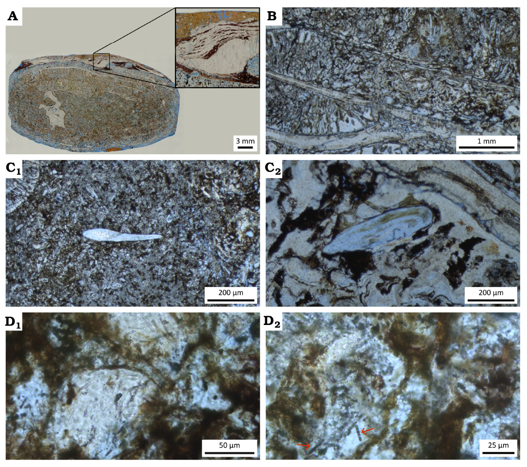
Fig. 1. Coprolite morphology and close-up of non-spherical inclusions in PV 634-P (thin section), shark coprolite from Coproland, Brazil, Rio do Rasto Formation, Guadalupian (middle Permian). A. Flattened heteropolar coprolite with interpreted biofilm laminae. B. Whorls exhibiting mucosal folds. C. Preserved fish scales showing internal structures (C1 ), 3-layered structure (C2 ). D. Filaments interpreted as filamentous cyanobacteria (D1 ), showing short, straight, non-branched structures with lighter sections consistent with cell-boundaries (D2 , cyanobacteria indicated byarrows).
In addition to spherical inclusions described below, several filamentous structures (up to 45) were observed in the thin section of the coprolite. These filaments are short, ranging 14–31 μm and are not branched (Fig. 1D2). They are very straight, with low flexibility. The filaments are translucent and lack coloration or mineral infilling. There are lighter sections that are consistent with the definition of cell boundaries, which appear to be rod-shaped.
The characteristics identified in the
filaments are very similar to those of the cyanobacteria
family Pseud-anabaenaceae. As seen in the studied material, the
Pseudana-baenaceae cyanobacteria have solitary trichomes, more or less
straight or a little flexuous (Komárek and
Johansen 2015). The cells are more or less spherical and lack
specialized cells such as heterocysts and akinetes (Komárek
and Johansen 2015) known from other cyanobacteria or
algae. This family is known to contribute to the
formation of microbial mats (McGregor and Rasmussen 2008;
Zammit et al. 2021). The microbial mats, in turn,
contribute to the fossil preservation by delaying
the decay of organic matter (Iniesto et al. 2016).
The presence of a spiral valve intestine that forms a heteropolar coprolite, mucosal folds and a piscivorous diet is indication of a shark producer (McAllister 1985; Dentzien-Dias et al. 2012; Rodrigues et al. 2018).
Spherical inclusions.—Two different kinds of larger spherical structures (morphotypes A1 and A2) between 31 and 51 μm in diameter were also identified in the same thin section (Table 1).
Table 1. Characteristics of egg types recovered from Coproland coprolites. L, length; W, width.
|
Coprolite |
Eggs |
Eggs morphology |
Eggs size (µm) |
Shell thickness (µm) |
Oncosphere (µm) |
|
UFRGS-PV-634-P (this study) |
20 per packet |
packet, rounded eggs (A1) |
32.4 –50.8 |
1.2–1.7 |
17.5–29.6 |
|
UFRGS-PV-634-P (this study) |
9 |
isolated, rounded eggs (A2) |
31.1–40.2 |
3.5–5.3 |
12.3–28.8 |
|
UFRGS-PV-429-P |
122 |
grouped, oval-shaped eggs with operculum |
L: 154.6–142.2 W: 97.7–84.6 |
2.4–4.3 |
L: 84.9–101.4 W: 44.1–74.3 |
A1: A first finding includes a cluster of at least 20 spherical structures surrounded by an envelope (Fig. 2A). The spherical structures have elliptical to sub-spherical shapes with diameters 32–51 μm (Table 1). The spherical structures within packets displayed a thin but distinct outer wall (1–2 μm thick) and in at least five spheres the presence of hook-like structures were confirmed. They are herein interpreted as a cluster of approximately 20 tightly packed eggs embedded within a probable gelatinous matrix. This relates to the following observations:
(i) Relative wall thickness is too big to be confused with arthropod eggs, spherical host cells or fungal spores which could be superficially confused with helminth eggs (Garcia et al. 2017).
(ii) Their internal structure is most consistent with tapeworm eggs having a thin but distinct outer shell, encapsulating an oncosphere (diameter 17–29 μm) with three pairs of hooks (Fig. 2B, a key characteristic of hexacanth eggs of modern eucestode tapeworms [Hoberg et al. 1997; Conn and Swiderski 2008; Sures et al. 2025]). In one egg cross section, structures seem to be repeated 3 times which would be consistent with three pair of hooks. In 4 other eggs similar structures are present although not necessarily as symmetrical or appearing 3 times which can be explained by different angles, position within the egg and location of where the egg was sliced during the thin section preparation (Fig. 3). The dimensions of these structures relative to oncosphere or outer shell is consistent with those reported from other eggs (Nasin et al. 1997: figs. 62, 64; Ivanov and Brooks 2002: fig. 22; De Chambrier 2006: fig. 20)
(iii) The arrangement including varying dimensions and occasional sub-spherical shapes resemble closely the arrangement as seen in cross sections of tapeworm egg packets (De Chambrier 2006). The size and morphology of these packets closely resemble those found in egg packets of modern tapeworms such as Dipylidium caninum (commonly found in canids; Pandey et al. 2022) and Kapsulotaenia (a cestode infecting reptiles and amphibians; De Chambrier 2006). According to Tyler (2006) four species of Echinobothrium (Cestoda: Diphyllidea), that parasitise the spiral valve of extant rays, have rounded eggs packaged in cocoons with 2–10 eggs. Calliobothrium can have 6–20 or even more than 23 oncospheres per cocoon (Nasin et al. 1997; Ivanov and Brooks 2002; Bernot 2015).
(iv) The arrangement of the eggs in packets (cocoons) suggests that they were originally released as a cohesive structure, a typical reproductive strategy among modern cestodes, where egg packets are shed in host faeces and dispersed in aquatic or terrestrial environments. The differences in size and shape of eggs (Fig. 2A) are consistent with sectioning such a packet (Nasin et al. 1997; De Chambrier 2006; Bernot et al. 2016).
A2: In another part of the thin section, there are several isolated spherical structures with a notably thicker outer shell (Fig. 4; Table 1). These eggs are slightly smaller in diameter than those included in clusters of A1, ranging 31–40 μm, which may indicate a possible variation in cestode species or differences in developmental stages. This relates to the following observations:
(i) Relative wall thickness of individual spherical structures is too big to be confused with arthropod eggs, spherical host cells or fungal spores or protozoan cysts which could be superficially confused with helminth eggs (Garcia et al. 2017).
(ii) The internal structures are consistent with: a prominent oncosphere, with some structure which may potentially be suggestive of hooks although they may also refer to other structures (Fig. 4A2). These structures, their relative size as well as their position straddling the oncosphere is reminiscent of hooks in a fully developed hexacanth within the egg (Monte et al. 2018). There are however also various other structures which could confirm to this morphology: relative size and position particularly during earlier stages of development like polar bodies, yolk mass, vitelline cell or macromeres (Ogren 1959, 1962), so the positive identification of these structures as hooks needs further confirmation. It is however consistent with the expectation that we would rather expect a fully developed hexacanth in shedded eggs rather than premature stages still in development. A distinct embryophore surrounding the oncosphere, structurally similar to that of extant taeniid cestodes (Chew 1983). An outer protective layer, which in some specimens exhibited signs of mineralization, possibly due to diagenetic processes.
(iii) Comparison to modern Helminths: The relative dimension of the layered structure as well as presence of oncospheres with one egg hinting at structures reminiscent of hooks with similar relative dimensions those represented in the hexacanth stage strongly indicates a eucestode tapeworm origin rather than a nematode or trematode (De Baets et al. 2021a). More detailed comparisons with modern forms in sharks are difficult as most stages are studied while still inside their hosts and more detailed information is mostly available for mammal or bird parasites. Furthermore, experimental taphonomy remains challenging.
Nevertheless, the similarity to eggs and strategies of modern taeniids suggests that these Permian parasites may have shared an ecological role with present-day tapeworms, relying on a multi-host life cycle involving both intermediate and definitive hosts.
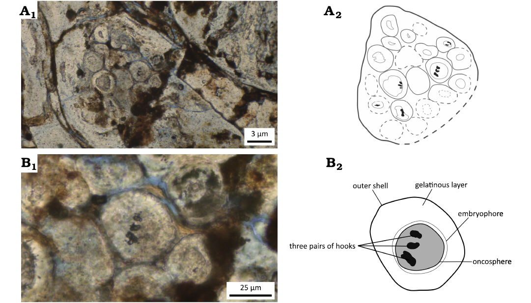
Fig. 2. Close-up of spherical inclusions type A1 in PV 634-P (thin section), shark coprolite from Coproland, Brazil, Rio do Rasto Formation, Guadalupian (middle Permian). A. Cluster of (sub)spherical bodies of type A1 interpreted as tapeworm eggs within an egg packet (cocoon). B. A single spherical body showing three pairs of hook-like structures and a distinct outer shell surrounded by others. A1, B1, photos with Leica DM750P petrographic microscope; A2, B2, interpretative drawings.
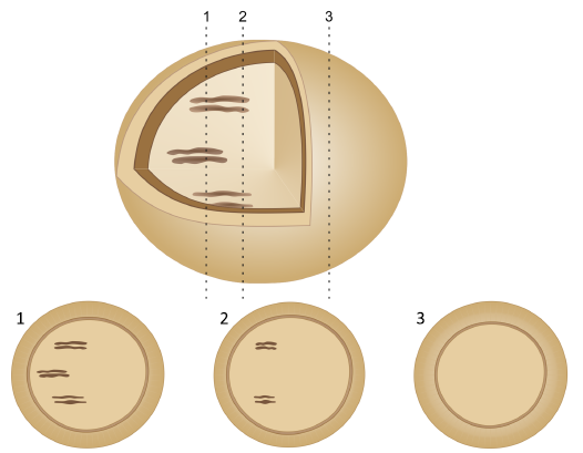
Fig. 3. Hypothetical cross sections through a more 3D-preserved egg and how it may effect the observable structures beyond other constraints like preservation.
Preservation features.—The eggs exhibit varying degrees of mineralization, with some showing authigenic mineral replacement of the outer shell also observed in other tapeworm eggs from another coprolite from the same locality (Dentzien-Dias et al. 2013). Certain eggs appear collapsed or distorted, suggesting minor diagenetic alteration. The coprolites display differential preservation, with some showing oxide caps that may have influenced the eggs’ fossilization. The eggs were evenly distributed throughout the coprolites, indicating they were likely shed in faeces rather than externally introduced. Over the coprolite a stack of fine laminae composed of organic matter (Fig. 1A) is deposited. The thickness of such stacks ranges 0.7–2 mm. The laminae are curved, and between them, lenses of the siltstone occur. The laminae are interpreted as a biofilm formed when the faeces were fresh.
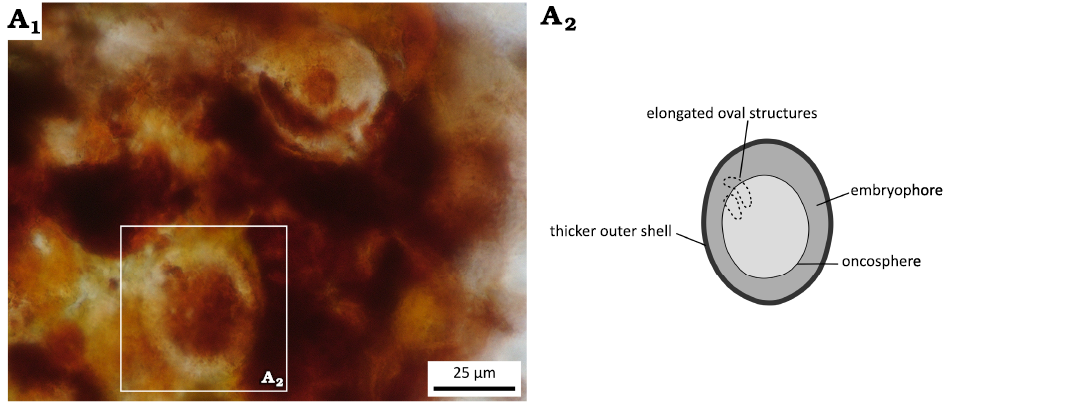
Fig. 4. Close-up of spherical inclusions type A2 in PV 634-P (thin section), shark coprolite from Coproland, Brazil, Rio do Rasto Formation, Guadalupian (middle Permian). Isolated spherical bodies interpreted as tapeworm eggs. A1, photo with Keyence HX-7000; A2, interpretative drawing.
Discussion
Affinity and evolutionary implications.—It is not always straightforward to separate intestinal parasitic remains from other microscopic inclusions, particularly fungal or plant remains (Szwabe and Kurnatowski 2012; Garcia et al. 2017). Some helminth eggs have been confused or are superficially similar to plant remains particularly cells or pollen (Garcia et al. 2017; Camacho et al. 2020). The spherical structures in the thin section of coprolite UFRGS-PV-634-P clearly differ from other inclusions based on their shape, size and associations and are not reminiscent of other known morphologies of host cells or microorganismal remains (Dentzien-Dias et al. 2017; Garcia et al. 2017; Dentzien-Dias and Łaska 2025) including fungal or other spores (Jurigan et al. 2023; Catafesta et al. 2026). In addition, pollen of angiosperms, which have the highest chances to be confused with tapeworm eggs (Szwabe and Kurnatowski 2012) are not yet present in Permian environments. Based on their internal structure and associations, the structures are most reminiscent of tapeworm eggs, including those previously reported from the same locality (Dentzien-Dias et al. 2013) as well as others with the Rio do Rasto Formation (Catafesta et al. 2026). The new findings include eggs that are smaller than previously described and associated in different arrangements, augmenting the diversity of preserved tapeworm-related structures at the Coproland locality from one to at least two, likely three, morphotypes.
The structures interpreted as a hooked oncosphere and egg packets strongly support a cestode (tapeworm) origin (De Baets et al. 2024). Hints at 3 paired structures in some of the eggs seems to corroborate such an interpretation (see Fig. 2B). The absence of these structures may have various explanations ranging from difference in preservation or developmental stage to more different position in thin sections (see Fig. 2B). The presence of egg packets (cocoons) suggests that the tapeworm species may have had a reproductive strategy similar to modern dipylidiid, echinobothriid or taeniid cestodes, which release egg packets in faeces (De Chambrier 2006; Tyler 2006; Pandey et al. 2022). The isolated eggs with thicker shells may represent a different cestode species or less likely an alternative developmental stage.
The co-occurrence of abundant eggs with two clearly distinct morphologies: (i) clustered eggs within probable proglottid fragments or mucosal packets, and (ii) isolated eggs with a thicker outer shell, within a single coprolite provides strong evidence of simultaneous infection by multiple cestode species. These two morphotypes differ in shell wall thickness, size, and distribution pattern (Table 1). Quite some variation in cocoon packaging and their response to contact with seawater (environment) has been observed even within the same genus or even same species of Callibothrium (Bernot 2015), but not as extreme differences in egg morphology as observed here. Both type of eggs provide examples of oncospheres with up to three pairs of hooks, confirming their interpretation as eucestode (true tapeworm) eggs (De Baets et al. 2021a, 2024). The clustered eggs suggest a reproductive strategy involving egg aggregation and passive shedding via proglottids or mucosal packaging. The clustered arrangement of eggs resembles egg packets known from modern echinobothriid (known from elasmobranchs) as well as dipylidiid or kapsulotaeniid cestodes (primarily described in terrestrial taxa). Although we deem it is less likely, such a structure could potentially also result from mucosal encapsulation or degeneration of reproductive tissue (e.g., proglottids) prior to fossilization. Extant echinobothriids have different morphology and packing of the eggs, having oval and rounded eggs organized as a single packets with 2–10 eggs or in chains (Tyler 2006). We also cannot entirely rule out that taphonomic processes such as compression or microbial stabilization may have enhanced the clustered appearance in the fossil record, although seems unlikely as structure of coprolite and their position is largely preserved and seems most likely to reflect differences in transmission strategies.
The second morphotype, isolated and with thicker shells, likely represents a separate species rather than a different developmental stage, as such variation in dispersal strategy and shell architecture is uncommon within a single cestode species. This supports the inference of multiple tapeworm taxa co-infecting the same definitive host (Curran and Caira 1995).
Additionally, a third morphotype previously described from another coprolite (Dentzien-Dias et al. 2013; De Baets et al. 2015a), attributed to the same type of elasmobranch producer and locality, comprises elongated proglottid fragments containing larger, more elliptical eggs, also bearing three pairs of hook-like structures. This further supports a broader cestode diversity with differing transmission strategies in the Coproland ecosystem (cf. Penadés-Suay et al. 2022).
Moreover, the presence of abundant
eggs with these distinct morphologies strongly supports the
interpretation that the coprolites derive from a definitive
host rather than an intermediate host that may have accidentally
ingested parasite eggs. Intermediate hosts typically carry larval stages
rather than shed large numbers of eggs from multiple adult cestode
species. These findings imply that cestode communities within Permian
aquatic hosts were more taxonomically
diverse than previously appreciated. The presence of distinct egg
morphologies and associations also raises the possibility of differing
life history strategies among the elasmobranch tapeworms, indicating the
establishment of different strategies already in the Palaeozoic.
Possible hosts and life cycle implications.—The presence of fish scales in the coprolites, mucosal folds and the heteropolar spiral morphology of the coprolite suggest that the final host was a shark, which served as the definitive host for the tapeworms (McAllister 1985; Dentzien-Dias et al. 2012; Rodrigues et al. 2018). The occurrence of two morphologically distinct egg types within a single thin section of the same coprolite points to simultaneous infection by multiple cestode species. This is consistent with modern elasmobranchs, which often harbour more than one adult tapeworm species in their spiral valve intestines and show differences in reproductive strategies (Curran and Caira 1995; Caira and Jensen 2001; Ivanov and Brooks 2002; Alarcos et al. 2006; Penadés-Suay et al. 2022). In modern tapeworm life cycles, the eggs are produced by adult cestodes in the final host and excreted in faeces. The clear presence of egg clusters and isolated eggs in the coprolite supports the conclusion that the eggs were produced by different adult cestodes rather than by intermediate stages or digestion byproducts. As with most modern cestodes, these Permian forms likely had complex life cycles involving at least two hosts (Scholz 1999; Leung 2021; Scholz and Kuchta 2022; Sures et al. 2025): a first intermediate host, likely a small aquatic invertebrate such as a crustacean, and a final vertebrate host (Fig. 5). Most analyses suggest an ancestral intermediate host to have been a crustacean and the majority of known modern tapeworm life cycles in aquatic environments involve a 3-host life cycle with the first intermediate host being a invertebrate and the final host being a vertebrate (Mackiewicz 1988; Sakanari and Moser 1989; Caira and Reyda 2005; Sures et al. 2025). Both 2-host or 3-host life cycles are observed in lineages parasitizing sharks as final hosts, but some have up to 2 additional paratenic hosts. The first intermediate is typically an invertebrate (mostly crustacean) and final host a vertebrate, although bony fishes could potentially also be intermediate hosts (Leung 2021). Ostracods and conchostracans are abundant in the Rio do Rasto Formation and constitute plausible first intermediate hosts (Azevedo et al. 2018). Bivalves are also common in the Rio do Rasto Formation but have so far not been found within this type of coprolite (Dentzien-Dias et al. 2012), so their function as hosts is unlikely. Bivalves are regularly reported from coprolites (Brachaniec et al. 2015) including Early Triassic freshwater ones (Yates et al. 2012) and some freshwater bivalves today can even survive passage through digestive system of fishes (Brown 2007) so their absence likely represents a true absence rather than a taphonomic bias. We cannot exclude other small invertebrates such as soft-bodied insect larvae that are not preserved or vertebrates (fish) as possible alternative intermediate hosts.
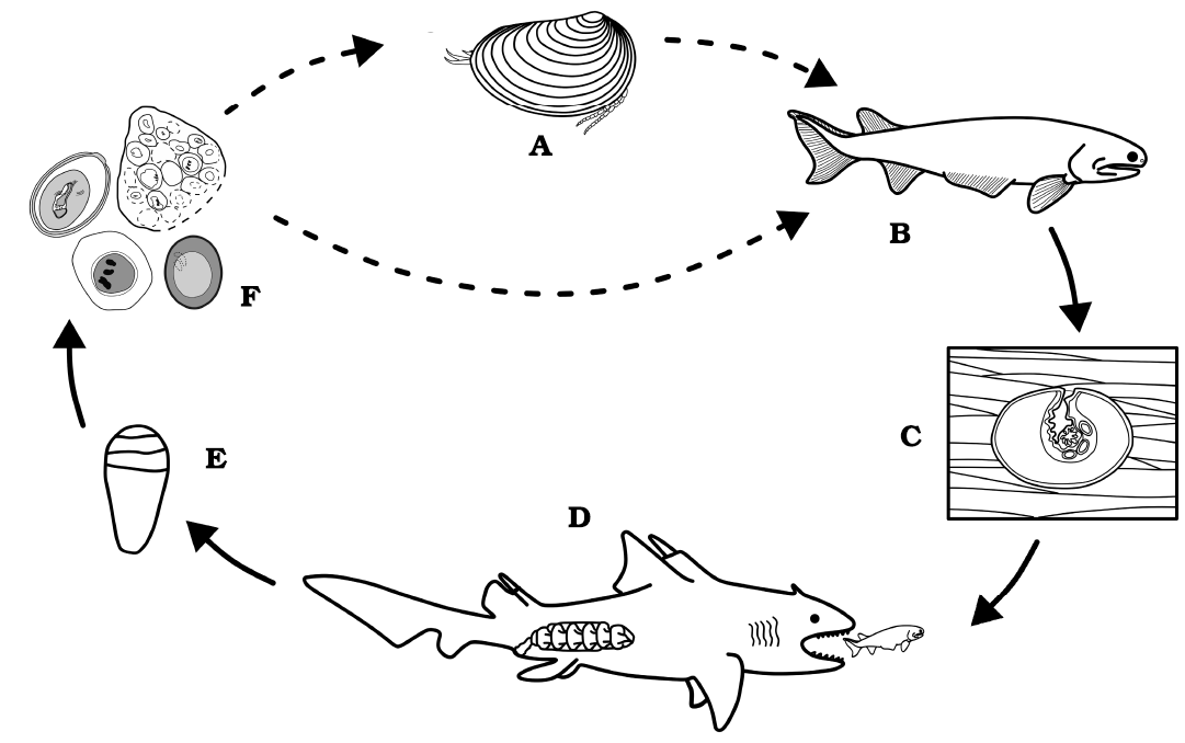
Fig. 5. Hypothetical reconstruction of life cycles of tapeworm parasites recovered from shark coprolites from Coproland, Brazil, Rio Do Rasto Formation, Guadalupian (middle Permian). Filled arrows are supported by direct evidence, stippled arrows are less certain, and possibilities suggested from circumstantial inference as discussed in text. A. Conchostracan as representative for a possible first (invertebrate) intermediate host. B. Bony fish as (2nd or 1st) intermediate host. C. Possible tapeworm egg development inside the host muscle tissue. D. Shark with spiral intestine as definitive host. E. Defecation. F. Tapeworm eggs found within the faeces (now preserved as mineralized coprolites).
A likely intermediate host are smaller fishes abundant in the Rio do Rasto ecosystems and represented in coprolites yielding tapeworm eggs by their scales. Shortened 2-host lifecycles cannot be ruled out (Leung 2021) but are considered unlikely and it is difficult to imagine the represented fishes to regularly feed on tapeworm propagules. Small fish as abundantly represented by their scales within the coprolite are therefore likely rather second intermediate than first (and only) intermediate hosts. The ingestion of this infected prey by an apex predator such as a shark would have assisted in completing the parasite’s life cycle. This trophic structure supports the concept of a “trophic transmission vacuum”, in which the incorporation of a second intermediate host bridges the ecological gap between microinvertebrates and large definitive hosts (Benesh et al. 2014).
There is no direct evidence for fishes preying on small crustaceans such as conchostracans and ostracods or reports of parasitized crustaceans in the Rio do Rasto Formation. However, remains of smaller fishes and small crustaceans often co-occur in fossiliferous beds (Azevedo et al. 2018), making such interactions plausible. While ostracods have not yet been found in association with definitive-host coprolites from this site, they have been found in coprolites attributed to palaeonisciform fishes from other Permian localities (Jurigan et al. 2023). Similarly, conchostracan remains have been documented in Permian coprolites assigned to tetrapods (Owocki et al. 2012; Niedźwiedzki et al. 2016; Sennikov et al. 2020), supporting their preservation potential in Permian vertebrate coprolites. These findings suggest that although such crustaceans have not yet been recovered from fish coprolites at the Rio do Rasto Formation, they could principally be found but seem absent.
Reconstructing the complex life cycles of helminths, particularly those of tapeworms infecting sharks, remains difficult and poorly understood, particularly in the absence of molecular approaches to assign their undifferentiated larval stages (Jensen and Bullard 2010; Randhawa 2011; Randhawa and Brickle 2011; Caira and Jensen 2014; Bennett et al. 2019, 2023; Scholz and Kuchta 2022). Jarecka (1961) noted that egg morphology can influence transmission to first intermediate hosts in freshwater systems, particularly crustaceans, although those studies focused on bird-definitive-host systems. More recently, Rasmussen and Randhawa (2018) demonstrated that diet plays a key role in shaping tapeworm diversity in sharks. These observations highlight the ecological complexity and evolutionary significance of trophic interactions in parasite life cycles. Reconstructing extinct parasite life cycles can be even more challenging. This is illustrated by attempts to reconstruct the life cycle of unknown parasites involved in the formation of blister pearls in Emsian ammonoid and bivalve molluscs (De Baets et al. 2011). It is tempting to invoke the presence of complex life cycles involving predatory gnathostome fishes as final hosts which prey on these molluscs as intermediate hosts given superficially similar pathologies are mostly observed in intermediate hosts of modern gymnophallid trematodes (De Baets et al. 2011, 2015b). However, parasitic remains preserved within the Devonian blister pearls in addition to inferred latter Cretaceous appearance of gymnophallid trematodes are not consistent with the attribution of these pathologies to flatworms (De Baets et al. 2015a; Huntley and De Baets 2015; Leung 2017, 2021). Lack of direct evidence for gnathostome parasitic infestations (Lukševics et al. 2009) or predator-prey relationships (e.g., in the form of coprolite inclusions) in these ecosystems further hamper the full reconstruction of these Devonian parasite life cycles.
With the available evidence from the Rio do Rasto Formation, we can hypothesize the following life cycles of cestode involving a Permian elasmobranch as the definitive host as follows: (i) eggs are released in the faeces of the definitive elasmobranch host and are likely ingested by a free-living, filter-feeding invertebrate; (ii) inside the invertebrate, the egg hatches, releasing a hexacanth larva, which penetrates the gut wall, encysts, and develops into a procercoid larva, typically in an organ such as the liver; (iii) the infected invertebrate is then consumed by a second host, a fish; (iv) the larva again penetrates the gut wall, encysts in the liver, and develops into a plerocercoid larva; (v) when the fish is eaten by the third and final host, the elasmobranch, the larva matures in the host’s spiral intestine; (vi) sexual reproduction occurs there, either through cross-fertilization or self-fertilization between adult worms, completing the cycle (Fig. 5). We have no direct fossil information to constrain the presence or identity of the 1st intermediate invertebrate host (hence this part of life cycle is more speculative), but inferring one seems more plausible than a 2-host life cycle just involving bony and cartilaginous fishes (Leung 2021) based on arguments discussed above. Further work is needed to corroborate this life cycle and more fully understand the food and interaction webs in the Rio do Rasto Formation.
Factors behind egg abundance.—The repeated discovery of cestode eggs in Permian bromalites strengthens evidence for early host-parasite coevolution in vertebrate digestive systems, complementing earlier findings from the Carboniferous of the USA (Zangerl and Case 1976). Bromalites is a term referring to any fossilized digestive matter originating from animals (Hunt and Lucas 2021) but so far tapeworm eggs have only been reported from Permian shark coprolites (fossilized faeces) and a Carboniferous cololite (fossilized intestinal tract contents preserved in situ). These findings suggest that complex parasitic life cycles of tapeworms were already well established in the Palaeozoic while evidence is lacking for parasitic nematodes such as ascaridoid nematodes. The abundance of eggs per bromalite attributable to tapeworms in Palaeozoic occurrences is high (29–122) with rare exceptions (Catafesta et al. 2026) compared to later Mesozoic and Cenozoic occurrences where pre-Quaternary reports typically lack tapeworm eggs or have putative isolated egg preserved if at all (De Baets et al. 2024). Preservational factors may therefore also play a crucial role and more studies on taphonomy of fossil coprolites are needed (Dentzien-Dias et al. 2018; Dentzien-Dias and Łaska 2025). Various factors could explain or work in concert to explain the high abundance and diversity of tapeworm eggs in coprolites.
Exceptional preservation (Qvarnström et al. 2016) may have played a role in the preservation of tapeworm eggs, particularly at the Coproland locality. The faecal matrix likely provided a protective microenvironment that minimized decay, facilitating the preservation of more delicate egg structures. Oxide-rich layers and microbial activity may have promoted early diagenetic mineralization within these particular coprolites, enhancing the durability of egg structures. The presence of authigenic minerals within the eggshell layers further suggests early diagenetic mineral replacement (Dentzien-Dias et al. 2013). Notably, 32 thin sections of different coprolites were examined, and cestode eggs were recovered in only two (ca. 6 %), including previous reports by Dentzien-Dias et al. (2013), suggesting an exceptional combination of preservational and biological factors. This is quite similar to reports from other studies of the frequency of helminth eggs (5–7 %) in fossil coprolites (Cardia et al. 2018; Barrios-de Pedro et al. 2020). In the two thin sections where eggs were present, they occurred in high abundance, highlighting that when conditions were favourable to their presence, these parasites were remarkably well represented. Biofilms on the surface of the coprolite indicates that an early mineralogical precipitation was likely biologically induced. This reinforces the primary role of microbial activity on coprolite preservation (Hollocher et al. 2001). According to Noffke et al. (2022), biofilms can provide a buffered microenvironment that enhances fossil preservation, especially in soft tissues and delicate structures such as helminth eggs. Microorganism-induced lithification has been shown to be a key mechanism in fossil preservation within coprolites (Hollocher and Hollocher 2012). Packaging eggs in packets or proglottids may also increase their preservation potential (Zamparo 2001; De Baets et al. 2024). However, the findings of abundant tapeworm eggs in Palaeozoic bromalites is not restricted to the Coproland locality and other post-Palaeozoic fossil coprolites yielding tapeworms have yielded mostly isolated eggs if at all.
Ecological factors might also
contribute to the high concentration of cestode eggs in
these fossils. It is possible that the Carboniferous–Permian aquatic
ecosystems more generally exhibited a high frequency of
parasite-host interactions, contributing to widespread
infection. The co-occurrence of abundant potential hosts, such as small
crustaceans and fishes, suggests that these ecosystems supported the
complex trophic relationships necessary for completing cestode life
cycles. The environment of the Rio Do Rasto Formation may
have the ecosystem particularly susceptible given its restricted nature
and occasional draughts (Azevedo et al. 2018; Francischini
et al. 2018b). The high number of coprolites found in only one
layer, with evidences of dry periods, might indicate that the producers
were occasionally restricted in one small area that may have intensified
the transmission (see Dentzien-Dias et al. 2012 for
discussion). Interestingly, all tapeworm remains from Rio do Rasto
Formation or Palaeozoic as a whole derive from shark
bromalites hinting the ecology may also be an important factor. The
spiral intestine is the most heavily parasitized internal organ of
modern elasmobranchs as it is the primary site occupied by cestodes.
It is fairly common for certain elasmobranchs to host
ten or more tapeworm species in their spiral intestines (Caira
and Healy 2004; Caira et al. 2012).
Therefore, individual conditions related to tapeworm transmission and their hosts may have influenced the concentration of eggs in specific coprolites. The studied coprolites may originate from highly infested individuals of particular hosts, thereby leading to a high shedding rates of eggs in faeces and skewing parasite abundance in the fossil record. Helminths frequently reproduce in higher-trophic level vertebrates that tend to be large and long-lived, which may generate a trophic transmission vacuum. Tapeworms may also have already used strategies to increase their transmission (Mackiewicz 1988). This transmission gap could be filled through incorporation of intermediate hosts, further supporting the likelihood that complex life cycles were already present in the Palaeozoic (Benesh et al. 2014, 2022; Parker et al. 2015). While the current dataset may hint at diverse strategies early in their evolution, further exceptional discoveries and sampling will be necessary to confirm the full complexity of cestode life cycles throughout the Phanerozoic. Mesozoic and (pre-Holocene) Cenozoic sampling has focused on particularly terrestrial tetrapods (Poinar and Boucot 2006; Hugot et al. 2014; Cardia et al. 2018, 2019, 2021; Dentzien-Dias et al. 2018; Francischini et al. 2018a; Barrios-de Pedro et al. 2020; Chin 2021; Oyarzún-Ruiz et al. 2021; Carmo et al. 2024). A possible tentacular structure in Cretaceous amber may represent the only potential fossil evidence for tapeworms in Mesozoic sharks (Luo et al. 2024) but its affinity, life cycle and taphonomic scenario needs further scrutiny (De Baets et al. 2024). The lack of ascaridoid nematode eggs in Palaeozoic is also notable given they have been mostly commonly reported from fossil coprolites in Mesozoic and Cenozoic usually attributed to their resistant eggs (Brinkkemper and van Haaster 2012; Dufour and Le Bailly 2013). It is, however, tempting to attribute this absence to their inferred origin in the tetrapods and the hypothesis of later colonization of elasmobranchs and teleost fishes (Li et al. 2018) but ecological, preservational and sampling factors discussed earlier may also explain lack of discoveries so far.
Conclusions
This study expands the fossil record of intestinal parasites, reporting new cestode eggs occurrences in Permian coprolites from the Coproland outcrop. The findings underscore the importance of fossilized faeces in reconstructing parasite-host interactions (Chin 2021) and suggest that intestinal tapeworms with complex life cycles were widespread among Permian sharks. The exceptional preservation and abundance of eggs is likely linked to a combination of ecological and taphonomic factors such as coprolite mineralization, bio-induced mineralization and oxide-rich sediments. These results confirm that intestinal tapeworm were already widespread among Permian sharks including forms already differing in reproductive strategies, supporting the idea about various forms with complex parasitic life cycles were well-established by the Palaeozoic and imply a long-term diversity in cestode reproductive strategies.
Authors’ contributions
PD-D and KDB designed and performed analyses and interpretations, wrote the draft manuscript; PD-D and LC made and described thin sections; ACLP, AS, and WL aided with interpretations. All co-authors read, contributed and agreed with final submitted manuscript.
Acknowledgements
We kindly thank Tim Littlewood (Natural History Museum, London, UK), an anonymous reviewer, and the editor for their constructive comments and suggestions which helped to further improve our manuscript. This study was funded by the National Science Centre (NCN), Poland, under the OPUS 27 grant (2024/53/B/NZ8/03528) to KDB. KDB, WŁ and AS were supported by the I.3.4 Action of the Excellence Initiative—Research University Programme at the University of Warsaw (Project: PARADIVE). This collaboration also benefitted from a research visit PD-D to Warsaw that was funded through Action I.1.1/IV.1.1 “Mentoring Programme” (to AS and KDB). KDB, AS and PD-D thank the Paleosynthesis Project and the Volkswagen Stiftung for funding the BITE workshop which benefitted this project (Az 96 796). PD-D thanks CNPq (Brazilian National Council of Scientific and Technological Development) for financing partially this research.
Editor: Andrzej Kaim
References
Alarcos, A., Ivanov, V., and Sardella, N. 2006. Distribution patterns and interactions of cestodes in the spiral intestine of the narrownose smooth-hound shark, Mustelus schmitti Springer, 1939 (Chondrichthyes, Carcharhiniformes). Acta Parasitologica 51: 100–106. Crossref
Azevedo, K.L., Schemiko, D.C.B., Soares, M.B., Vega, C.S., and Vesely, F.F. 2018. The São Jerônimo da Serra Site, Rio do Rasto Formation (Middle/Upper Permian), Paraná Basin, Brazil: faciological and taphonomic context. Brazilian Journal of Geology 48: 821–837. Crossref
Barrios-de Pedro, S., Osuna, A., and Buscalioni, Á.D. 2020. Helminth eggs from early cretaceous faeces. Scientific Reports 10: 18747. Crossref
Benesh, D.P., Chubb, J.C. and Parker, G.A. 2014. The trophic vacuum and the evolution of complex life cycles in trophically transmitted helminths. Proceedings of the Royal Society B: Biological Sciences 281 (1793): 20141462. Crossref
Benesh, D.P., Chubb, J.C., Lafferty, K.D., and Parker, G.A. 2022. Complex life-cycles in trophically transmitted helminths: Do the benefits of increased growth and transmission outweigh generalism and complexity costs? Current Research in Parasitology & Vector-Borne Diseases 2: 100085. Crossref
Bennett, J., Jorge, F., Poulin, R., and Randhawa, H. 2019. Revealing trophic transmission pathways of marine tapeworms. Parasitology Research 118: 1435–1444. Crossref
Bennett, J., Presswell, B., and Poulin, R. 2023. Tracking life cycles of parasites across a broad taxonomic scale in a marine ecosystem. International Journal for Parasitology 53: 285–303. Crossref
Bernot, J.P. 2015. Taxonomy, Systematics, and Host Associations of Cestodes of Triakid Sharks. 152 pp. Master thesis, University of Connecticut, Storrs.
Bernot, J.P., Caira, J.N., and Pickering, M. 2016. Diversity, phylogenetic relationships and host associations of Calliobothrium and Symcallio (Cestoda : ‘Tetraphyllidea’) parasitising triakid sharks. Invertebrate Systematics 30: 616–634. Crossref
Boos, A.D.S., Schultz, C.L., Vega, C.S., and Aumond, J.J. 2013. On the presence of the Late Permian dicynodont Endothiodon in Brazil. Palaeontology 56: 837–848. Crossref
Brachaniec, T., Niedźwiedzki, R., Surmik, D., Krzykawski, T., Szopa, K., Gorzelak, P., and Salamon, M.A. 2015. Coprolites of marine vertebrate predators from the Lower Triassic of southern Poland. Palaeogeography, Palaeoclimatology, Palaeoecology 435: 118–126. Crossref
Brinkkemper, O. and van Haaster, H. 2012. Eggs of intestinal parasites whipworm (Trichuris) and mawworm (Ascaris): Non-pollen palynomorphs in archaeological samples. Review of Palaeobotany and Palynology 186: 16–21. Crossref
Brown, R.J. 2007. Freshwater mollusks survive fish gut passage. Arctic 60: 124–128. Crossref
Bueno, V.M. and Caira, J.N. 2023. Phylogenetic relationships, host associations, and three new species of a poorly known group of “tetraphyllidean” tapeworms from elasmobranchs. Zootaxa 5254: 30–50. Crossref
Caira, J.N. and Healy, C.J. 2004. Elasmobranchs as hosts of metazoan parasites. In: J.C. Carrier, J.A. Musick, and M.R. Heithaus (eds.), Biology of Sharks and Their Relatives, 523–551. CRC Press, Boca Raton. Crossref
Caira, J.N. and Jensen, K. 2001. An investigation of the co-evolutionary relationships between onchobothriid tapeworms and their elasmobranch hosts. International Journal for Parasitology 31: 960–975. Crossref
Caira, J.N. and Reyda, F.B. 2005. Eucestoda (true tapeworms). In: K. Rohde (ed.), Marine Parasitology, 92–104. CSIRO Publishing, Collingwood.
Caira, J.N. and Jensen, K. 2014. A Digest of Elasmobranch Tapeworms. Journal of Parasitology 100: 373–391. Crossref
Caira, J.N., Healy, C.J., and Jensen, K. 2012. An updated look at elasmobranchs as hosts of metazoan parasites. In: J.C. Carrier, J.A. Musick and M.R. Heithaus (eds.), Biology of Sharks and Their Relatives, 547–578. CRC Press, Boca Raton. Crossref
Caira, J.N., Jensen, K., and Healy, C.J. 1999. On the phylogenetic relationships among tetraphyllidean, lecanicephalidean and diphyllidean tapeworm genera. Systematic Parasitology 42: 77–151. Crossref
Camacho, M., Perri, A., and Reinhard, K. 2020. Parasite microremains: preservation, recovery, processing, and identification. In: A.G. Henry (ed.), Handbook for the Analysis of Micro-Particles in Archaeological Samples, 173–199. Springer International Publishing, Cham. Crossref
Campbell, R.A. 1983. Parasitism in the deep sea. In: G.T. Rowe (ed.), The Sea, 473–552. John Wiley & Sons, New York.
Cardia, D.F., Bertini, R.J., Camossi, L.G., and Letizio, L.A. 2018. The first record of Ascaridoidea eggs discovered in Crocodyliformes hosts from the Upper Cretaceous of Brazil. Revista Brasileira de Paleontologia 21: 238–244. Crossref
Cardia, D.F., Bertini, R.J., Camossi, L.G., and Letizio, L.A. 2019. Two new species of ascaridoid nematodes in Brazilian Crocodylomorpha from the Upper Cretaceous. Parasitology International 72: 101947. Crossref
Cardia, D.F., Bertini, R.J., Camossi, L.G., Richini-Pereira, V.B., Losnak, D.O., Francischini, H., and Dentzien-Dias, P. 2021. Paleoparasitological analysis of a coprolite assigned to a carnivoran mammal from the Upper Pleistocene Touro Passo Formation, Rio Grande do Sul, Brazil. Anais da Academia Brasileira de Ciencias 93 (Supplement 2): e20190876. Crossref
Carmo, G.M. do, Luiz, A.H. da S.G., Passos, J.F., Lima, S. de S., Araújo-Júnior, H.I. de, and Pereira, F.B. 2024. First report of a morulated Ascaridoidea (Nematoda) egg in an avian coprolite from the Paleogene of the Paraíba Valley, State of São Paulo, Brazil. Journal of Helminthology 98: e60. Crossref
Catafesta, L.P., Dentzien-Dias, P., Calaça, F.J.S., Dias, E.V., Łaska, W., De Baets, K., and Francischini, H. 2026. Paleoecological insights from coprolites and their inclusions from the Permian Rio do Rasto Formation, Brazil. Palaeoworld 35: 201060. Crossref
Chew, M.W.K. 1983. Taenia crassiceps ultrastructural observations on the oncosphere and associated structures. Journal of Helminthology 57: 101–113. Crossref
Chin, K. 2021. Gastrointestinal parasites of ancient nonhuman vertebrates: evidence from coprolites and other materials. In: K. De Baets and J.W. Huntley (eds.), The Evolution and Fossil Record of Parasitism: Coevolution and Paleoparasitological Techniques, 359–375. Springer, Cham. Crossref
Cisneros, J.C., Dentzien-Dias, P., and Francischini, H. 2021. The Brazilian pareiasaur revisited. Frontiers in Ecology and Evolution 9: 758802. Crossref
Conn, D.B. and Swiderski, Z. 2008. A standardised terminology of the embryonic envelopes and associated developmental stages of tapeworms (Platyhelminthes: Cestoda). Folia Parasitologica 55: 42–52. Crossref
Curran, S. and Caira, J.N. 1995. Attachment site specificity and the tapeworm assemblage in the spiral intestine of the blue shark (Prionace glauca). The Journal of Parasitology 81: 149–157. Crossref
Da Silva, P.A., Borba, V.H., Dutra, J.M., Leles, D., Da-Rosa, A.A., Ferreira, L.F., and Araujo, A. 2014. A new ascarid species in cynodont coprolite dated of 240 million years. Anais da Academia Brasileira de Ciências 86: 265–270. Crossref
De Chambrier, A. 2006. Redescription of Kapsulotaenia sandgroundi (Carter, 1943) (Eucestoda: Proteocephalidea: Acanthotaeniinae), a parasite of Varanus komodoensis (Varanoidea: Varanidae) from Indonesia. Systematic Parasitology 63: 81–91. Crossref
De Baets, K. and Littlewood, D.T.J. 2015. The importance of fossils in understanding the evolution of parasites and their vectors. Advances in Parasitology 90: 1–51. Crossref
De Baets, K., Dentzien-Dias, P., Harrison, G.W.M., Littlewood, D.T.J. and Parry, L.A. 2021a. Fossil constraints on the timescale of parasitic helminth evolution. In: K. De Baets and J.W. Huntley (eds.), The Evolution and Fossil Record of Parasitism: Identification and Macroevolution of Parasites, 231–271. Springer, Cham. Crossref
De Baets, K., Dentzien-Dias, P., Huntley, J.W., Vanhove, M.P.M., Łaska, W., Skawina, A., Steenkiste, N.W.L.V., and Vanadzina, K. 2024. Fossil constraints on the origin and evolution of Platyhelminthes are surprisingly concordant with modern molecular phylogenies. Zoologia (Curitiba) 41: e24002. Crossref
De Baets, K., Dentzien-Dias, P., Upeniece, I., Verneau, O., and Donoghue, P.C. 2015a. Constraining the deep origin of parasitic flatworms and host-interactions with fossil evidence. Advances in Parasitology 90: 93–135. Crossref
De Baets, K., Huntley, J.W., Klompmaker, A.A., Schiffbauer, J.D. and Muscente, A.D. 2021a. The fossil record of parasitism: its extent and taphonomic constraints. In: K. De Baets and J.W. Huntley (eds.), The Evolution and Fossil Record of Parasitism: Coevolution and Paleoparasitological Techniques, 1–50. Springer, Cham. Crossref
De Baets, K., Keupp, H. and Klug, C. 2015a. Parasites of ammonoids. In: C. Klug, D. Korn, K. De Baets, I. Kruta, and R.H. Mapes (eds.), Ammonoid Paleobiology: From Anatomy to Ecology, 837–875. Springer, Dordrecht. Crossref
De Baets, K., Klug, C., and Korn, D. 2011. Devonian pearls and ammonoid-endoparasite co-evolution. Acta Palaeontologica Polonica 56: 159–180. Crossref
Dentzien-Dias, P. and Łaska, W. 2025. Analyzing coprolites: Different methods of the study of coprolites. In: S.G. Lucas, A.P. Hunt, and H. Klein (eds.), Vertebrate Ichnology: Fish Ichnology, Consumption, Burrows and Reproduction, Geoconservation, 283–297. Elsevier, Cham. Crossref
Dentzien-Dias, P.C., Carrillo-Briceño, J.D., Francischini, H., and Sánchez, R. 2018. Paleoecological and taphonomical aspects of the Late Miocene vertebrate coprolites (Urumaco Formation) of Venezuela. Palaeogeography, Palaeoclimatology, Palaeoecology 490: 590–603. Crossref
Dentzien-Dias, P.C., de Figueiredo, A.E.Q., Horn, B., Cisneros, J.C., and Schultz, C.L. 2012. Paleobiology of a unique vertebrate coprolites concentration from Rio do Rasto Formation (Middle/Upper Permian), Paraná Basin, Brazil. Journal of South American Earth Sciences 40: 53–62. Crossref
Dentzien-Dias, P.C., Poinar Jr., G., and Francischini, H. 2017. A new actinomycete from a Guadalupian vertebrate coprolite from Brazil. Historical Biology 29: 770–776. Crossref
Dentzien-Dias, P.C., Poinar Jr, G., de Figueiredo, A.E.Q., Pacheco, A.C.L., Horn, B.L., and Schultz, C.L. 2013. Tapeworm eggs in a 270 million-year-old shark coprolite. PLoS One 8 (1): e55007. Crossref
Dufour, B. and Le Bailly, M. 2013. Testing new parasite egg extraction methods in paleoparasitology and an attempt at quantification. International Journal of Paleopathology 3: 199–203. Crossref
Fontanelli, R.C.O. and Vega, C.S. 2020. Coprólitos da Formação Rio do Rasto (Neopermiano da Bacia do Paraná) registrados no Estado do Paraná. Boletim Paranaense de Geociências 77 (3): 20–25. Crossref
Francischini, H., Dentzien-Dias, P., and Schultz, C.L. 2018a. A fresh look at ancient dungs: Brazilian Triassic coprolites revisited. Lethaia 51: 389–405. Crossref
Francischini, H., Dentzien-Dias, P., Guerra-Sommer, M., Menegat, R., Santos, J.O.S., Manfroi, J., and Schultz, C.L. 2018b. A middle Permian (Roadian) lungfish aestivation burrow from the Rio do Rasto Formation (Paraná Basin, Brazil) and associated U-Pb dating. Palaios 33: 69–84. Crossref
Garcia, L.S., Arrowood, M., Kokoskin, E., Paltridge, G. P., Pillai, D. R., Procop, G. W., Ryan, N., Shimizu, R. Y., and Visvesvara, G. 2017. Practical guidance for clinical microbiology laboratories: laboratory diagnosis of parasites from the gastrointestinal tract. Clinical Microbiology Reviews 31 (1): e00025-17. Crossref
Gonçalves, M.L.C., Araújo, A., and Ferreira, L.F. 2003. Human intestinal parasites in the past: new findings and a review. Memórias do Instituto Oswaldo Cruz 98: 103–118. Crossref
Hoberg, E.P., Mariaux, J., Justine, J.-L., Brooks, D.R., and Weekes, P.J. 1997. Phylogeny of the orders of the Eucestoda (Cercomeromorphae) based on comparative morphology: historical perspectives and a new working hypothesis. The Journal of Parasitology 83: 1128–1147. Crossref
Hollocher, K.T. and Hollocher, T.C. 2012. Early processes in the fossilization of terrestrial feces to coprolites, and microstructure preservation. New Mexico Museum of Natural History and Science Bulletin 57: 79–91.
Hollocher, T.C., Chin, K., Hollocher, K.T., and Kruge, M.A. 2001. Bacterial residues in coprolite of herbivorous dinosaurs: role of bacteria in mineralization of feces. Palaios 16: 547–565. Crossref
Hugot, J.-P., Gardner, S.L., Borba, V., Araujo, P., Leles, D., Stock Da-Rosa, Á.A., Dutra, J., Ferreira, L.F., and Araújo, A. 2014. Discovery of a 240 million year old nematode parasite egg in a cynodont coprolite sheds light on the early origin of pinworms in vertebrates. Parasites & Vectors 7: 486. Crossref
Hunt, A.P. and Lucas, S.G. 2021. The ichnology of vertebrate consumption: dentalites, gastroliths and bromalites. New Mexico Museum of Natural History & Science Bulletin 87: 1–136.
Hunt, A.P. and Lucas, S.G. 2025. Consumulites. In: S.G. Lucas, A.P. Hunt, and H. Klein (eds.), Vertebrate Ichnology: Fish Ichnology, Consumption, Burrows and Reproduction, Geoconservation, 405–452. Elsevier, Amsterdam. Crossref
Huntley, J.W. and De Baets, K. 2015. Trace fossil evidence of trematode, bivalve parasite, host interactions in deep time. Advances in Parasitology 90: 201–231. Crossref
Iniesto, M., Buscalioni, Á.D., Carmen Guerrero, M., Benzerara, K., Moreira, D., and López-Archilla, A.I. 2016. Involvement of microbial mats in early fossilization by decay delay and formation of impressions and replicas of vertebrates and invertebrates. Scientific Reports 6: 25716. Crossref
Ivanov, V.A. and Brooks, D.R. 2002. Calliobothrium spp. (Eucestoda: Tetraphyllidea: Onchobothriidae) in Mustelus schmitti (Chondrichthyes: Carcharhiniformes) from Argentina and Uruguay. Journal of Parasitology 88: 1200–1213. Crossref
Jarecka, L. 1961. Morphological adaptations of tapeworm eggs and their importance in the life cycles. Acta Parasitologica Polonica 9: 409–426.
Jensen, K. and Bullard, S.A. 2010. Characterization of a diversity of tetraphyllidean and rhinebothriidean cestode larval types, with comments on host associations and life cycles. International Journal for Parasitology 40: 889–910. Crossref
Jensen, K., Mojica, K.R., and Caira, J.N. 2014. A new genus and two new species of lecanicephalidean tapeworms from the striped panray, Zanobatus schoenleinii (Rhinopristiformes: Zanobatidae), off Senegal. Folia Parasitologica 61: 432–440. Crossref
Jurigan, I., Ricardi-Branco, F., and Dentzien-Dias, P. 2023. Permian Western Gondwana food chain elucidated by coprolites from the Corumbataí Formation (Paraná Basin, Brazil). Journal of South American Earth Sciences 127: 104414. Crossref
Komárek, J. and Johansen, J.R. 2015. Filamentous Cyanobacteria. In: J.D. Wehr, R.G. Sheathand, and J.P. Kociolek (eds.), Freshwater Algae of North America. Second Edition, 135–235. Academic Press, Boston. Crossref
Leung, T.L. 2017. Fossils of parasites: what can the fossil record tell us about the evolution of parasitism? Biological Reviews 92: 410–430. Crossref
Leung, T.L. 2021. Parasites of fossil vertebrates: What we know and what can we expect from the fossil record? In: K. De Baets and J.W. Huntley (eds.), The Evolution and Fossil Record of Parasitism: Identification and Macroevolution of Parasites, 1–27. Springer, Cham. Crossref
Li, L., Lü, L., Nadler, S.A., Gibson, D.I., Zhang, L.-P., Chen, H.-X., Zhao, W.-T., and Guo, Y.-N. 2018. Molecular phylogeny and dating reveal a terrestrial origin in the early Carboniferous for ascaridoid nematodes. Systematic Biology 67: 888–900. Crossref
Lukševics, E., Lebedev, O., Mark-Kurik, E., and Karatajūtė-Talimaa, V. 2009. The earliest evidence of host–parasite interactions in vertebrates. Acta Zoologica 90 (s1): 335–343. Crossref
Luo, C., Palm, H.W., Zhuang, Y., Jarzembowski, E.A., Nyunt, T.T., and Wang, B. 2024. Exceptional preservation of a marine tapeworm tentacle in Cretaceous amber. Geology 52: 497–501. Crossref
Mackiewicz, J.S. 1988. Cestode transmission patterns. The Journal of Parasitology 74: 60–71. Crossref
McAllister, J.A. 1985. Reevaluation of the formation of spiral coprolites. The University of Kansas, Paleontological Contributions 114: 1–12.
McGregor, G.B. and Rasmussen, J.P. 2008. Cyanobacterial composition of microbial mats from an Australian thermal spring: a polyphasic evaluation. FEMS Microbiology Ecology 63: 23–35. Crossref
Monte, G.L.S., Cavalcante, D.G., and Oliveira, J.B.S. 2018. Parasitic profiling of Japanese quails (Coturnix japonica) on two farms with conventional production system in the Amazon region. Pesquisa Veterinária Brasileira 38: 847–851. Crossref
Nasin, C.S., Caira, J.N., and Euzet, L. 1997. Analysis of Calliobothrium (Tetraphyllidea: Onchobothriidae) with descriptions of three new species and erection of a new genus. The Journal of Parasitology 83: 714–733. Crossref
Niedźwiedzki, G., Bajdek, P., Qvarnström, M., Sulej, T., Sennikov, A.G., and Golubev, V.K. 2016. Reduction of vertebrate coprolite diversity associated with the end-Permian extinction event in Vyazniki region, European Russia. Palaeogeography, Palaeoclimatology, Palaeoecology 450: 77–90. Crossref
Noffke, N., Mángano, M.G., and Buatois, L.A. 2022. Biofilm harvesters in coastal settings of the early Palaeozoic. Lethaia 55: 1–18. Crossref
Nonsrirach, T., Morand, S., Ribas, A., Manitkoon, S., Lauprasert, K., and Claude, J. 2023. First discovery of parasite eggs in a vertebrate coprolite of the Late Triassic in Thailand. PLOS One 18 (8): e0287891. Crossref
Ogren, R.E. 1959. The hexacanth embryo of a dilepidid tapeworm. The formation of shell and inner capsule around the oncosphere. The Journal of Parasitology 45: 580–585. Crossref
Ogren, R.E. 1962. Embryonic development of a dilepidid tapeworm. Transactions of the American Microscopical Society 81: 65–72. Crossref
Owocki, K., Niedźwiedzki, G., Sennikov, A.G., Golubev, V.K., Janiszewska, K., and Sulej, T. 2012. Upper Permian vertebrate coprolites from Vyazniki and Gorokhovets, Vyatkian Regional Stage, Russian Platform. Palaios 27: 867–877. Crossref
Oyarzún-Ruiz, P., Pérez-Espinoza, S.A., González-Saldías, F., Martin, F., and Moreno, L. 2021. Paleoparasitological survey of coprolites of Darwin’s ground sloth Mylodon darwini (Xenarthra, Folivora: Mylodontidae) from Cueva del Milodón Natural Monument, Chilean Patagonia. Archaeological and Anthropological Sciences 13: 138. Crossref
Pandey, R.P., Raj, V.S., Singh, R.K., and Mohaptara, T.M. 2022. Dipylidiasis. In: S.C. Parija and A. Chaudhury (eds.), Textbook of Parasitic Zoonoses, 377–383. Springer Nature, Singapore. Crossref
Parker, G.A., Ball, M.A., and Chubb, J.C. 2015. Evolution of complex life cycles in trophically transmitted helminths. Host incorporation and trophic ascent. Journal of Evolutionary Biology 28: 267–291. Crossref
Penadés-Suay, J., Jarque-Rico, A.E., Tomás, J., and Aznar, F.J. 2022. Determinants of diversity and composition of the tapeworm fauna of blue sharks, Prionace glauca: a geographical and host-specificity analysis. Journal of Helminthology 96: e87. Crossref
Poinar, Jr., G. and Boucot, A.J. 2006. Evidence of intestinal parasites of dinosaurs. Parasitology 133: 245–249. Crossref
Qvarnström, M., Niedźwiedzki, G., and Žigaitė, Ž. 2016. Vertebrate coprolites (fossil faeces): an underexplored Konservat-Lagerstätte. Earth-Science Reviews 162: 44–57. Crossref
Randhawa, H.S. 2011. Insights using a molecular approach into the life cycle of a tapeworm infecting great white sharks. Journal of Parasitology 97: 275–280. Crossref
Randhawa, H.S. and Brickle, P. 2011. Larval parasite gene sequence data reveal cryptic trophic links in life cycles of porbeagle shark tapeworms. Marine Ecology Progress Series 431: 215–222. Crossref
Randhawa, H.S. and Poulin, R. 2010. Determinants of tapeworm species richness in elasmobranch fishes: untangling environmental and phylogenetic influences. Ecography 33: 866–877. Crossref
Rasmussen, T.K. and Randhawa, H.S. 2018. Host diet influences parasite diversity: a case study looking at tapeworm diversity among sharks. Marine Ecology Progress Series 605: 1–16. Crossref
Rocha-Campos, A.C., Basei, M.A.S., Nutman, A.P., Santos, P.R., Passarelli, C.R., Canile, F.M., Rosa, O.C.R., Fernandes, M.T. , Santa Ana, H., and Veroslavsky, G. 2019. U-Pb zircon dating of ash fall deposits from the Paleozoic Paraná Basin of Brazil and Uruguay: a reevaluation of the stratigraphic correlations. The Journal of Geology 127: 167–182. Crossref
Rodrigues, M.I.C., da Silva, J.H., Santos, F.E.P., Dentzien-Dias, P., Cisneros, J.C., de Menezes, A.S., Freire, P.T.C., and Viana, B.C. 2018. Physicochemical analysis of Permian coprolites from Brazil. Spectrochimica Acta Part A: Molecular and Biomolecular Spectroscopy 189: 93–99. Crossref
Sakanari, J.A. and Moser, M. 1989. Complete life cycle of the elasmobranch cestode, Lacistorhynchus dollfusi Beveridge and Sakanari, 1987 (Trypanorhyncha). The Journal of Parasitology 75: 806–808. Crossref
Scholz, T. 1999. Life cycles of species of Proteocephalus, parasites of fishes in the Palearctic Region: a review. Journal of Helminthology 73: 1–19. Crossref
Scholz, T. and Kuchta, R. 2022. Fish tapeworms (Cestoda) in the molecular era: achievements, gaps and prospects. Parasitology 149: 1876–1893. Crossref
Sennikov, A.G., Bulanov, V.V., and Scholze, F. 2020. Coprolite with conchostracans from the terminal Permian of Central Russia—paleobiological and stratigraphic significance. Paleontological Journal 54: 6–13. Crossref
Sianto, L., Chame, M., Silva, C.S., Gonçalves, M.L., Reinhard, K., Fugassa, M., and Araújo, A. 2009. Animal helminths in human archaeological remains: a review of zoonoses in the past. Revista do Instituto de Medicina Tropical de São Paulo 51: 119–130. Crossref
Sures, B., Díaz-Morales, D.M., Yong, R.Q.-Y., Erasmus, A., and Schwelm, J. 2025. Biology and life cycle of helminths. In: N.J. Smit and B. Sures (eds.), Aquatic Parasitology: Ecological and Environmental Concepts and Implications of Marine and Freshwater Parasites, 89–123. Springer Nature Switzerland, Cham. Crossref
Szwabe, K. and Kurnatowski, P. 2012. Comparative analysis of morphometric features of the eggs of selected alimentary tract parasites and of the plant pollens. Annals of Parasitology 58: 87–96.
Tyler, G.A.I. 2006. Tapeworms of elasmobranchs. A monograph on the Diphyllidea (Platyhelminthes, Cestoda). Bulletin of the University of Nebraska State Museum 20: 1–142.
Yates, A.M., Neumann, F.H., and Hancox, P.J. 2012. The earliest post-Paleozoic freshwater bivalves preserved in coprolites from the Karoo Basin, South Africa. PLOS One 7 (2): e30228. Crossref
Zammit, G., Schembri, S., and Fenech, M. 2021. Phototrophic biofilms and microbial mats from the marine littoral of the central Mediterranean. Acta Botanica Croatica 80: 112–120. Crossref
Zamparo, D. 2001. Phylogenetic Systematic Analysis of the Neodermata (Platyhelminthes) and Aspidobothrea (Trematoda: Neodermata) with Investigation of the Evolution of the Quinone Tanned Eggshell. 224 pp. MSc. Thesis, University of Toronto, Toronto.
Zangerl, R. and Case, G.R. 1976. Cobelodus aculeatus (Cope) an anacanthous shark from Pennsylvanian black shales of North America. Palaeontographica Abteilung A 154 (4–6): 107–157.
Acta Palaeontol. Pol. 71 (2): 323–335, 2026
http://doi.org/10.4202/app.01294.2025